ROS-ID?活性氧/活性氮检测试剂盒
自由基和其他活性种在许多心理和病理生理过程中发挥着重要作用。自由基一旦在细胞内产生,就会破坏多种细胞成分,包括蛋白质、脂质和DNA。然而,在较低浓度下,自由基可以作为细胞信号传导中的第二信使。
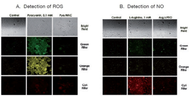
Enzo Life Sciences的ROS-ID??ROS/RNS detection kit可用荧光显微镜直接实时监测活细胞中活性氧与活性氮(ROS/RNS)的产生。该试剂盒中包含三种检测探针:NO检测试剂(红色,Ex/Em 650/670 nm),用于总ROS检测的氧化应激检测试剂(绿色,Ex/Em 490/525 nm)和超氧化物检测试剂(橙色,Ex/Em 550/620 nm)。试剂盒内还包括阳性对照,绿脓菌素和L-精氨酸,它们分别是ROS和NO产生的常见诱导剂;阴性对照N-乙酰-L-半胱氨酸和c-PTIO则分别是ROS和NO的常见清除剂。通过将三种检测探针与一组特异性抑制剂、激活剂相结合,能够区分超氧化物、一氧化氮和过氧亚硝酸盐。
产品特点
● 直接监测活细胞中的活性氧和氮化物(ROS/RNS)
●?可区分超氧化物、一氧化氮和过氧亚硝酸盐
●?高灵敏度、特异性和准确性,可用于活细胞研究
●?与组织培养基的主要成分(酚红、FBS和BSA)兼容
●?试剂盒内包含整套试剂,包括ROS/RNS诱导剂和清除剂
实验示例
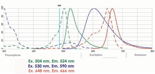
图1. 氧化应激检测试剂(绿色)、超氧化物检测试剂(蓝色)和NO检测试剂(红色)的激发和发射光谱。
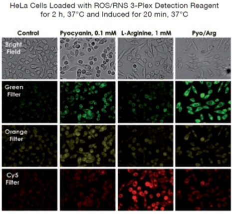
图2. 使用ROS/RNS检测试剂盒演示HeLa细胞中过氧亚硝酸盐和NO生成的代表性实验。分析表明,由于NO与超氧化物的反应,L-精氨酸和绿脓菌素的联合处理产生过氧亚硝酸盐(绿色荧光),但很少产生NO(红色荧光)。
??
产品信息
| 产品货号 | ENZ-51001-200 |
| 产品名称 | ROS-ID??ROS/RNS detection kit / 欣博盛生物 |
| 别名 | Reactive oxygen species / Reactive nitrogen species |
| 规格 | 1*1Kit |
| 长期保存 | -80°C |
| 试剂盒组分 | NO Detection Reagent (Red), 60μl Oxidative Stress Detection Reagent (Green), 300 nmoles Superoxide Detection Reagent (Orange), 300 nmoles NO Inducer (L-Arginine), 100 μl ROS Inducer (Pyocyanin), 1 μmole NO Scavenger (c-PTIO), 400 nmoles ROS Inhibitor (N-acetyl-L-cysteine), 2x10 mg 10X Wash Buffer, 15 ml |
| 应用 | Fluorescence microscopy |
部分产品引用文献
1.?Hypoxia-induced oxidative stress promotes therapy resistance via upregulation of heme oxygenase-1 in multiple myeloma: K. Abe, et al.; Cancer Med. (2023)
2.?Dihydroartemisinin-transferrin adducts enhance TRAIL-induced apoptosis in triple-negative breast cancer in a P53-independent and ROS-dependent manner: X. Zhou, et al.; Front. Oncol. 11, 789336 (2022)
3.?Intermittent exposure of cultured endothelial cells to physiologically relevant fructose concentrations has a profound impact on nitric oxide production and bioenergetics: M.L. Fiorello, et al.; PLoS One 17, e0267675 (2022)
4.?S1P (Sphingosine-1-Phosphate)-Induced Vasodilation in Human Resistance Arterioles During Health and Disease: B. Katunaric, et al.; Hypertension 79, 2250 (2022)
5.?Extra-mitochondrial citrate synthase initiates calcium oscillation and suppresses age-dependent sperm dysfunction: W. Kang, et al.; Lab. Invest. 100, 583 (2020)
6.?Liposome-encapsulated bacillus calmette-guérin cell wall skeleton enhances antitumor efficiency for bladder cancer in vitro and in vivo via induction of amp-activated protein kinase: Y.M. Whang, et al.; Cancers (Basel) 12, 3679 (2020)
7.?Mesenchymal stem cell conditioned medium ameliorates diabetic serum-induced insulin resistance in 3T3-L1 cells: A. Sanap, et al.; Chronic Dis. Transl. Med. 7, 47 (2020)
8.?Isoorientin derived from Gentiana veitchiorum Hemsl. flowers inhibits melanogenesis by down-regulating MITF-induced tyrosinase expression: Q. Wu, et al.; Phytomedicine 57, 129 (2019)
9.?Low intensity ultrasound reduces high glucose reduced nitric oxide generation in retinal pigment epithelial cells: M.B. Karmacharya, et al.; Ultrasound Med. Biol. 44, 647 (2018)
10.?HEK-293 secretome attenuates kainic acid neurotoxicity through insulin like growth factor-phosphatidylinositol-3-kinases pathway and by temporal regulation of antioxidant defense machineries: C. Venugopal, et al.; Neurotoxicology 69, 189 (2017)
11.?Selective enhancing effect of metal ions on mutagenicity: N. Fujii, et al.; Genes Environ. 38, 21 (2016)
12.?Cell-to-cell diffusion of glucose in the mammalian heart is disrupted by high glucose. Implications for the diabetic heart: W.C. De Mello ; Exp. Cell Res. 334, 239 (2015)
13.?Increased Proinflammatory Cytokine Production and Decreased Cholesterol Efflux Due to Downregulation of ABCG1 in Macrophages Exposed to Indoxyl Sulfate: K. Matsuo, et al. ; Toxins (Basel) 7, 3155 (2015)
14.?Regulation of gene expression by tobacco product preparations in cultured human dermal fibroblasts: G.E. Malpass, et al.; Toxicol. Appl. Pharmacol. 279, 211 (2014)
15.?Enterococcus faecalis infection causes inflammation, intracellular oxphos-independent ROS production, and DNA damage in human gastric cancer cells: J.A. Strickertsson, et al.; PLoS One 8, e63147 (2013)
本文来自互联网用户投稿,该文观点仅代表作者本人,不代表本站立场。本站仅提供信息存储空间服务,不拥有所有权,不承担相关法律责任。 如若内容造成侵权/违法违规/事实不符,请联系我的编程经验分享网邮箱:chenni525@qq.com进行投诉反馈,一经查实,立即删除!
- Python教程
- 深入理解 MySQL 中的 HAVING 关键字和聚合函数
- Qt之QChar编码(1)
- MyBatis入门基础篇
- 用Python脚本实现FFmpeg批量转换
- Python设计模式
- 基于多目标粒子群算法的支配解求解,基于多目标粒子群的帕累托前沿求解,基于mopso的多目标求解,基于多目标粒子群的模糊优化算
- 【Kubernetes 】Kubernetes 安全审计实战指南
- 【 C语言 】 | C程序百例
- 框架基础-网络编程+Tomcat服务器+XML
- 计算机网络-甘晴void学习笔记
- drf阶段测试题
- 从零开始做题:逆向 ret2text level0
- AI大概不会很快抢走你的饭碗哦!
- 地域与文化,智能酒精壁炉选择的多元因素