肺癌单细胞文献阅读1
写在前面
看点肺癌生信文献吧,只写点不了解的部分和觉得我认为有必要的部分,其余略过
文献
Single-cell RNA sequencing reveals distinct tumor
microenvironmental patterns in lung adenocarcinoma
IF:8.0 中科院分区:1区 医学 WOS分区:Q1
结论
1.heterogeneous carcinoma cell transcriptomes reflecting histological grade and oncogenic pathway activities, and two distinct
microenvironmental patterns
2.
3.
introduction
癌由恶性上皮细胞、非恶性免疫细胞 、非恶性基质细胞。
result
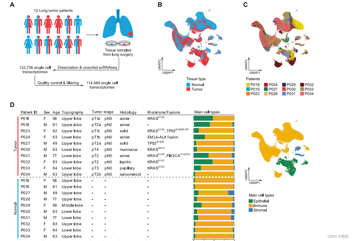
1.肺腺癌的细胞多样性
b、c看数据有没有去除批次效应,d看临床和病理信息,和分为上皮、免疫、基质细胞的比例关系,bd一起看,可看到癌样本大多是上皮细胞,且上皮细胞的比例占比差异还很大。正常大多为免疫细胞。

2.看肿瘤上皮细胞概况
1.只提取上皮细胞,看肿瘤纯度>90%,根据拷贝数变异,拷贝数方法:For copy-number assignment, InferCNV v1.3.3 was used with default parameters.

2.做了原癌基因的通路活性,涉及EGFR, TGFβ, JAK/STAT, Hypoxia, and
PI3K signaling。基因突变与通路之间的表征:
p53 signaling was significantly reduced in tumors harboring TP53 mutations,whereas pathway activity scores for EGFR and MAPK signaling were not significantly higher in KRAS-mutated compared to
KRAS-wildtype tumors。
方法:Oncogenic signaling pathway activity scores were computed using the R package “progeny”。
3.umap维度设置为低维度时,发现以组织类型聚类的趋势。
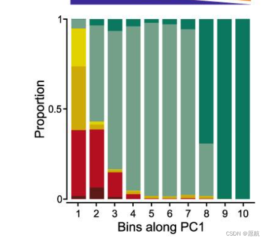
4.有与肺癌进展相关的基因,与PC1相关。SCGB3A1 and SCGB3A2
5.stroml与basal区别在哪?
6.pc1 in 10bins 啥意思?

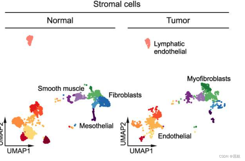
3.看基质细胞
1.纤维原细胞向纤维肌肉细胞转化

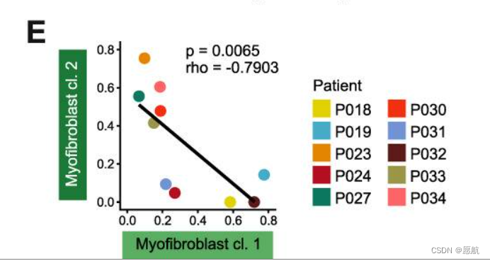
2.讲一堆纤维肌肉细胞亚型,亚型1仅在肿瘤中出现;亚型2在肿瘤与正常都出现,该亚型高表达胶原、基质降解酶、基质蛋白质,这说明了肿瘤中胞外基质重塑。亚型二高表达TGFβ and JAK/STAT signaling 、hypoxia-induced pathways,亚型一恰恰相反。二者细胞量在样本内呈反比。We
conclude that myofibroblasts cluster 1 and 2 represent “normal-like”
and “cancer-associated” phenotypes of myofibroblasts, respectively,
and either of them can predominate the stromal microenvironment
所以,这边应该算是定义了两个新亚型,并经实验验证。
4.看免疫细胞
1.看哪些?
monocyte-derived macrophages, monocytes, myeloid and plasmacytoid dendritic cells, mast cells, and T, NK, B,
and plasma cells
2.dendritic cells、myeloid cell增加,macrophages and monocytes减少。
3. monocyte-derived macrophage clusters 1 and 2,两亚群。
cluster 1 高表达与M2极化相关的 SELENOP, 跟免疫相关的通路低表达,。while cluster 2 高表达抗炎因子, such as CXCL9 and
CXCL10, the proinflammatory cytokine IL1B, and gene signatures related to immune response and M1 polarization。
4.再讲一下其他细胞在正常与肿瘤之间的分布, CD8+ T, B, and
plasma cells were increased, while NK and conventional T cells
were decreased。Regulatory T cells只在肿瘤中出现,表达CTLA4 and
TIGIT,呈免疫抑制功能。
5.两种模式的肺腺癌肿瘤微环境
找由细胞组(主要由哪几种细胞)构成的病人,区分根据定义为不同组,说明不同组存在的生存差异等信息。并做受体配体作用,最后经由tcga验证分组的生存与突变负荷等。
后结
第一篇看完的单细胞文章,感觉是先分大类,说明样本大类的区别,而后再根据其他文献对大类进行细分,找不同组别间小类的细胞组成区别,最后同时呈现,比较明显,再根据生物学去讲组别存在的差异可能蕴含的合理性。分析完大类及小类后,再重新将样本分类,观察分类后的样本的差异情况,并通过bulk数据验证。相比胃癌那篇,这篇比较难看懂,大多是生物学知识和背景。
本文来自互联网用户投稿,该文观点仅代表作者本人,不代表本站立场。本站仅提供信息存储空间服务,不拥有所有权,不承担相关法律责任。 如若内容造成侵权/违法违规/事实不符,请联系我的编程经验分享网邮箱:chenni525@qq.com进行投诉反馈,一经查实,立即删除!
- Python教程
- 深入理解 MySQL 中的 HAVING 关键字和聚合函数
- Qt之QChar编码(1)
- MyBatis入门基础篇
- 用Python脚本实现FFmpeg批量转换
- iMessage群发系统的功能设计和代码应用!
- uniapp 常用定时器实现方式
- C++高级编程——STL:list容器、set容器和map容器
- 天气预报网站windy的使用简介
- MSVS C# Matlab的混合编程系列2 - 构建一个复杂(含多个M文件)的动态库:
- 【Element】el-tree-select 限制只能同级选择
- 从零学Java List集合
- xshell (XFTP下载)免安装
- 1.1.0 IGP高级特性之BFD
- python零基础入门教程(非常详细),从零基础入门到精通,看完这一篇就够了